Anatomy Pictures Of Lower Back And Hip / Back Muscles And Low Back Pain - Muscle anatomy leg back female hamstring illustration joint anatomical buttocks fitness gracilis human musculature 3d illustration 3d rendering adductor magnus arthritis biceps femoris body calf muscle diagram gastrocnemius.. Adduction leg moves back towards midline. Browse 4,815 hip anatomy stock photos and images available, or search for hip replacement or knee anatomy to find more great stock photos and pictures. Pain that originates elsewhere may radiate to. The sacroiliac (si) joints connect the sacrum at the base of the spine with the hip bone. Up to 90% of people recover from sciatica without surgery.
Bones of the pelvis and lower back. Understanding the anatomy of your lower spine can help you communicate more effectively with the medical professionals who treat your lower back pain. When a person experiences lower back and hip pain simultaneously, there may be an underlying injury or medical. Cerebellum, cheek, cheeks, chin, dimples, ear, earache. Bones of the pelvis and lower back.
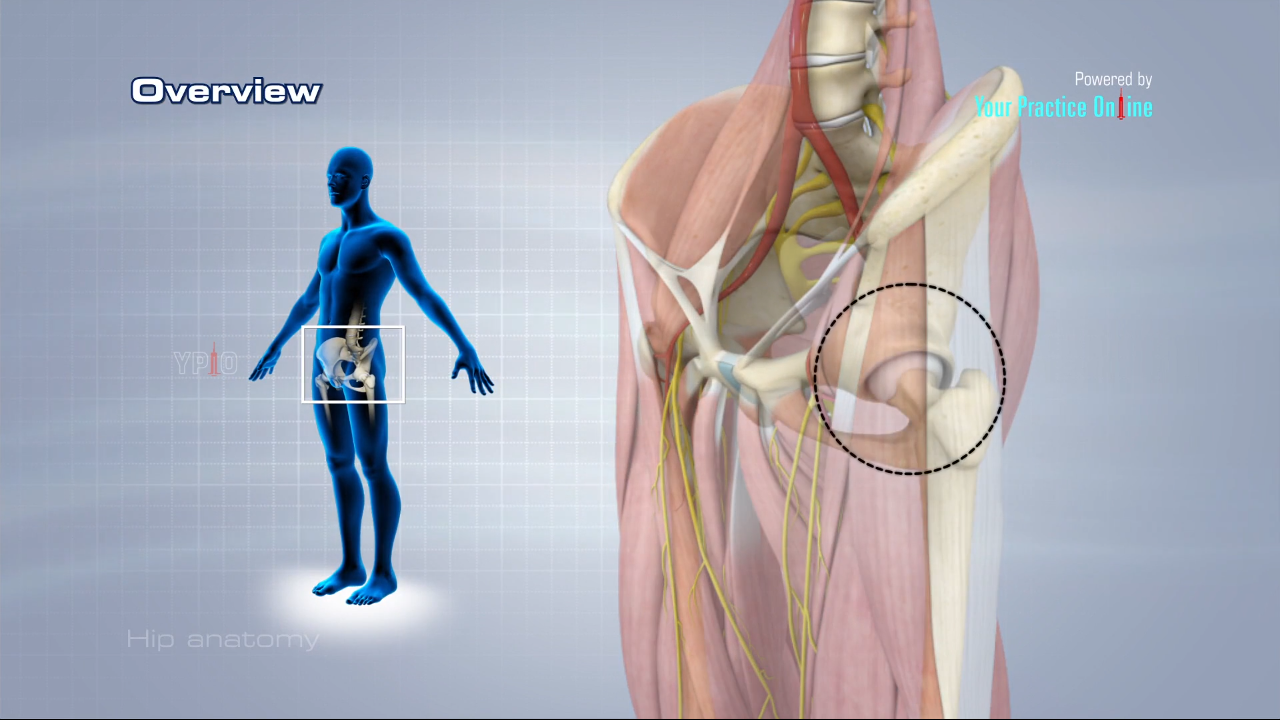
Hip Anatomy Video Hip Orthopaedics Videos Your Practice Online Education from www.ypo.education Adduction leg moves back towards midline. They provide a great deal of strength to modulate powerful forces between the upper and lower body. Other pelvic muscles, such as the psoas major and iliacus, serve as flexors of the trunk and thigh at the hip joint and. The bones of the pelvis and lower back work together to support the body's weight, anchor the abdominal and hip muscles, and protect the delicate vital organs of the vertebral and abdominopelvic cavities. Muscle strain is another common cause. Some of the other muscles in the hip are. The foot swings forward and comes back into contact with the floor with a heel strike active hip flexion. Browse 222 lower back skeleton stock photos and images available, or start a new search to explore more stock photos and images.
Understanding the anatomy of your lower spine can help you communicate more effectively with the medical professionals who treat your lower back pain.
Browse 222 lower back skeleton stock photos and images available, or start a new search to explore more stock photos and images. The fibers converge and pass posterolateral and upward, to form a tendon that runs across the back of the neck of the and is inserted into the trochanteric fossa of the femur. Hip pain may result from inflammation, degeneration, or injury to structures and tissues within the hip joint. Mri of upper leg (femur). The lumbar region of the spine, more commonly known as the lower back, is situated between the thoracic, or chest, region of the spine, and the sacrum. Up to 90% of people recover from sciatica without surgery. Most of the time, pain in your hip and groin is caused by a problem with the bones or other structures in or around the hip joint. A patient's guide to hip anatomy. It is the muscles of the hip that allow the movements of the hip. Adduction leg moves back towards midline. Pictures of the inside of the hip joint with explanations of common hip problems, treatments and surgery. The vertebral column of the lower back includes the five lumbar vertebrae, the sacrum, and the coccyx. Karir para artis march 20, 2021 groin, inguinal region and the fascia / aponeurosis:
Muscle anatomy leg back female hamstring illustration joint anatomical buttocks fitness gracilis human musculature 3d illustration 3d rendering adductor magnus arthritis biceps femoris body calf muscle diagram gastrocnemius. They provide a great deal of strength to modulate powerful forces between the upper and lower body. These muscles, including the gluteus maximus and the hamstrings, extend the thigh at the hip in support of the body's weight and propulsion. These sections are cervical (neck), thoracic (upper and middle back), lumbar (lower back), and sacrum (tailbone). 12 photos of the muscles of the lower back and buttocks diagram.
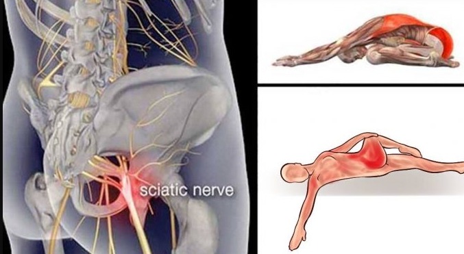
10 Piriformis Stretches To Get Rid Of Sciatica Hip And Lower Back Pain 01 Easy Life from 01easylife.com Hip pain may result from inflammation, degeneration, or injury to structures and tissues within the hip joint. The anatomy of the hip and back is comprised of numerous parts that can be injured or wear out, and many problems that occur in this area can display the exact same symptoms or pathology. Anatomy of the piriformis muscle. Some of the other muscles in the hip are. Adduction leg moves back towards midline. Anatomy of the human body. These sections are cervical (neck), thoracic (upper and middle back), lumbar (lower back), and sacrum (tailbone). The foot swings forward and comes back into contact with the floor with a heel strike active hip flexion.
Mri of upper leg (femur).
The different anatomical areas of the gluteal region: The sacroiliac (si) joints connect the sacrum at the base of the spine with the hip bone. The bones of the pelvis and lower back work together to support the body's weight, anchor the abdominal and hip muscles, and protect the delicate vital organs of the vertebral and abdominopelvic cavities. 12 photos of the muscles of the lower back and hip diagram. Understanding the anatomy of your lower spine can help you communicate more effectively with the medical professionals who treat your lower back pain. Understanding lower back anatomy is key to understanding the root of lower back and hip pain. The piriformis is a small muscle located deep within the hip and buttocks region. Knee feels weak despite exercise: Hip ad and flexion n: Normally, a smooth cushion of shiny white hyaline (or articular) cartilage about 1/4 inch thick covers the femoral head and the acetabulum.the articular cartilage is kept slick by fluid made in the synovial membrane (joint lining). When something injures or puts pressure on the sciatic nerve, it can cause pain in the lower back that spreads to the hip, buttocks, and leg. Browse 222 lower back skeleton stock photos and images available, or start a new search to explore more stock photos and images. The anatomy of the hip and back is comprised of numerous parts that can be injured or wear out, and many problems that occur in this area can display the exact same symptoms or pathology.
Mri of upper leg (femur). Karir para artis march 20, 2021 groin, inguinal region and the fascia / aponeurosis: Browse 222 lower back skeleton stock photos and images available, or start a new search to explore more stock photos and images. Some of the other muscles in the hip are. Pain that originates elsewhere may radiate to.
Smrt Hips Lower Back Abdomen Massage Magazine from www.massagemag.com Normally, a smooth cushion of shiny white hyaline (or articular) cartilage about 1/4 inch thick covers the femoral head and the acetabulum.the articular cartilage is kept slick by fluid made in the synovial membrane (joint lining). The back comprises the spine and spinal nerves, as well as several different muscle groups. Knee feels weak despite exercise: It is the muscles of the hip that allow the movements of the hip. The vertebral column of the lower back includes the five lumbar vertebrae, the sacrum, and the coccyx. Hip anatomy, function and common problems front view of the hip joint bones. Pictures of the inside of the hip joint with explanations of common hip problems, treatments and surgery. Bones of the pelvis and lower back.
Hip anatomy, function and common problems front view of the hip joint bones.
Normally, a smooth cushion of shiny white hyaline (or articular) cartilage about 1/4 inch thick covers the femoral head and the acetabulum.the articular cartilage is kept slick by fluid made in the synovial membrane (joint lining). Anatomy of the human body. Bones of the pelvis and lower back. It allows for complete rotations of the hip and is also. These sections are cervical (neck), thoracic (upper and middle back), lumbar (lower back), and sacrum (tailbone). Muscles of the lower back and hip diagram, human muscles, muscles of the lower back and hip diagram. Anatomy pictures of lower back and hip : Upper limb anatomy arm anatomy muscle anatomy anatomy study body anatomy anatomy thigh the muscles of the hip and thigh keep your hip joints strong and mighty, allowing for a wide range of hip movements. This anatomical atlas was especially designed for a specific public (radiologists. Browse 222 lower back skeleton stock photos and images available, or start a new search to explore more stock photos and images. To put it plainly, sometimes hip pain comes from the hip, but a lot of times hip pain comes from the back. Understanding lower back anatomy is key to understanding the root of lower back and hip pain. The hip joint is the uppermost part of the leg where the head of the thigh bone (femur) fits into the socket of the pelvis.